


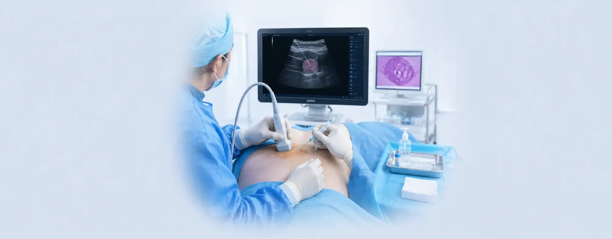
Hearing that you need a biopsy can feel daunting. For many people, the word itself raises concerns about pain, surgery, or what the results might reveal. In reality, most biopsies today are far less invasive than people expect. Advances in medical imaging now allow doctors to collect tissue samples with great precision, often through a small needle and without major surgery.
One such technique is an image-guided biopsy, where imaging technology helps doctors locate the exact area that needs to be sampled. This improves accuracy while minimising impact on the surrounding healthy tissue.
What Is a Biopsy?
A biopsy involves removing a small sample of tissue from the body for examination under a microscope. A specialist called a pathologist analyses the sample to help determine the cause of an abnormal finding, such as a lump, swelling, or an unusual change seen on a scan.
A biopsy doesn't automatically mean cancer is suspected. It may also be used to diagnose infections, inflammatory conditions, autoimmune diseases, or other medical disorders.
What Is an Image-Guided Biopsy?
An image-guided biopsy is a biopsy performed with the help of medical imaging. It is commonly performed as part of Interventional Radiology, where doctors use real-time images to guide the biopsy needle to the exact area that needs to be sampled — which is especially valuable when the abnormal area is small, deep, or cannot be felt by hand.
Different imaging techniques may be used depending on where the tissue is located:
| Imaging used | Uses radiation? | Often used for |
|---|---|---|
| Ultrasound | No | Breast, thyroid, lymph nodes, superficial masses |
| CT | Yes, low dose | Deep organs such as the lung, liver, or kidney |
| MRI | No | Lesions best seen only on MRI, e.g., some breast or prostate findings |
| Mammography (stereotactic) | Yes, low dose | Breast calcifications not visible on ultrasound |
When Is an Image-Guided Biopsy Recommended?
Your doctor may recommend an image-guided biopsy when an abnormal area cannot be easily felt during a physical examination, or when greater precision is needed. It may be used to investigate:
- Breast lumps detected on imaging
- Thyroid nodules
- Liver abnormalities
- Kidney lesions
- Lung nodules
- Enlarged lymph nodes
- Soft tissue masses
The goal is to obtain a tissue sample from the correct location while avoiding nearby blood vessels, nerves, and other important structures.
What Is a Needle Biopsy?
The most common type of image-guided biopsy is a needle biopsy. A thin or hollow needle is inserted through the skin to collect a small tissue sample; the specific needle used depends on the area being examined and how much tissue is needed.
Because only a small skin puncture is required, a needle biopsy is generally less invasive than a surgical biopsy and often allows for a quicker recovery.
How Does Image Guidance Work in Practice?
The imaging method chosen depends on where the abnormality is and how well it can be seen with each technique.
Ultrasound-Guided Biopsy
Ultrasound uses sound waves to create real-time images of the area being examined. As the doctor watches these images on a monitor, they guide the biopsy needle into the target tissue. Ultrasound guidance is commonly used for breast lumps, thyroid nodules, enlarged lymph nodes, some liver abnormalities, and superficial soft tissue masses. It provides continuous imaging throughout the procedure and does not use radiation, which is why it's often the preferred option when the target can be seen clearly with sound waves.
CT-Guided Biopsy
CT-guided biopsy uses cross-sectional X-ray imaging to pinpoint targets that are deeper in the body or surrounded by bone or air, such as lung nodules, some liver or kidney lesions, and masses deep in the abdomen or pelvis. The scanner takes images between needle adjustments so the doctor can confirm the needle's position before the sample is collected. It involves a low dose of radiation, which your care team will weigh against the diagnostic benefit.
MRI-Guided Biopsy
MRI-guided biopsy is used less often, generally reserved for lesions that are only visible on MRI and not on ultrasound, mammography, or CT — for example, certain breast or prostate findings. It doesn't use radiation, but the equipment and set-up are more specialised, so this procedure is typically performed at centres with dedicated MRI-biopsy capability.
Stereotactic (Mammography-Guided) Biopsy
For some breast abnormalities — particularly tiny calcium deposits (microcalcifications) that show up on a mammogram but aren't visible on ultrasound — a stereotactic biopsy uses mammography images taken from different angles to calculate the exact location of the target before the needle is inserted.
How to Prepare for Your Biopsy
Preparation varies depending on the type of biopsy and the area being examined, but your healthcare team will typically ask you to:
- Tell them about any blood-thinning medications or supplements you take, as these may need to be paused beforehand
- Mention any allergies, particularly to local anaesthetic or contrast dye
- Let them know if you are or might be pregnant
- Follow any fasting instructions if sedation will be used
- Arrange for someone to drive you home if sedation is planned
Always follow the specific instructions given by your healthcare provider, as these can vary between facilities and procedures.
What Happens During the Procedure?
Although the exact steps depend on the part of the body being examined, most image-guided biopsies follow a similar process:
- The skin is cleaned to reduce the risk of infection.
- A local anaesthetic is given to numb the area.
- Imaging is used to identify the target tissue.
- The biopsy needle is carefully guided into position.
- One or more tissue samples are collected.
- A small dressing is applied once the procedure is complete.
Many biopsies are completed within 30 to 60 minutes, and most patients are able to go home the same day.
Is the Procedure Painful?
This is one of the most common concerns patients have. Most people feel a brief sting when the local anaesthetic is injected. After that, you may notice some pressure during the procedure, but significant pain is uncommon.
It's also normal to have mild soreness or bruising for a day or two afterwards. Your healthcare team will explain how to care for the biopsy site and when you can return to your usual activities.
What Are the Risks?
Image-guided biopsies are generally very safe, and serious complications are uncommon. As with any procedure that breaks the skin, possible risks include:
- Bleeding or bruising at the biopsy site
- Infection
- Discomfort or soreness for a day or two
Some biopsy sites carry additional, procedure-specific risks — for example, a lung biopsy carries a small risk of a partial lung collapse (pneumothorax), which your team will monitor for after the procedure. Your doctor will explain the risks that are relevant to your specific biopsy and how they will be minimised and managed.
Getting Your Results
After the sample is collected, it's sent to a laboratory where a pathologist examines the tissue under a microscope, sometimes alongside specialised tests. This process usually takes several days to about a week, though timing can vary depending on the complexity of the testing required. Your doctor will discuss the results with you and explain what they mean for your care.
Are There Different Types of Biopsy Procedures?
Yes. There are several types of biopsy procedures, and the most appropriate one depends on the location of the abnormality, the amount of tissue needed, and the information your doctor is trying to obtain. Examples include:
- Needle biopsy
- Surgical biopsy
- Endoscopic biopsy
- Bone marrow biopsy
Whenever appropriate, doctors often prefer minimally invasive techniques such as image-guided needle biopsies, as they usually involve a smaller incision, a shorter recovery, and fewer complications than surgery.
Conclusion
An image-guided biopsy is a precise, minimally invasive way of obtaining tissue samples for diagnosis. By using imaging such as ultrasound, CT, or MRI to guide the biopsy needle, doctors can accurately target the area of concern while limiting the impact on surrounding healthy tissue.
Whether you're having a needle biopsy or an ultrasound-guided biopsy, understanding the procedure can make the experience feel less intimidating. If you have questions before your appointment, don't hesitate to raise them with your healthcare team — they can explain why the biopsy has been recommended and what to expect on the day.
How PB Health Can Support You
We understand that undergoing a biopsy can feel overwhelming. At PB Health, our radiologists, pathologists, and clinical teams work together to ensure every biopsy procedure is performed with precision, safety, and compassion. From advanced image-guided techniques to clear explanations of your results, we're here to support you through every stage of your diagnosis and care.
This article is for general information only and is not a substitute for professional medical advice. Please speak with your doctor about what to expect from your specific procedure.